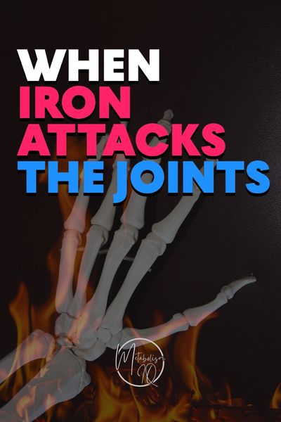
Arthritis: A Metabolic View of Degeneration
Arthritis is often described as “wear and tear,” but that view misses the deeper truth. Joints don’t simply erode with age — they break down metabolically. Beneath every ache and stiffness lies a web of disrupted cellular communication, oxidative stress, and iron imbalance. When redox systems collapse, cartilage loses its elasticity, synovial membranes inflame, and bone remodeling spins out of control. Arthritis, in this sense, isn’t a localized joint problem — it’s a reflection of the body’s internal chemistry. To restore mobility and comfort, we must begin not with the joint itself, but with the cellular metabolism that sustains it.
The Cellular Landscape of a Joint
A joint isn’t just bone and cartilage. It’s a living, breathing ecosystem — a microenvironment of highly specialized cells that work together to maintain structure, lubrication, immunity, and repair. When this cellular orchestra plays in balance, movement is fluid and pain-free. But when iron builds up and redox balance collapses, inflammation, stiffness, and degeneration take hold.
Chondrocytes — The Cartilage Builders
Chondrocytes are the architects of articular cartilage, the smooth tissue that allows bones to glide effortlessly against one another. These cells produce collagen type II and proteoglycans like aggrecan, which give cartilage its bounce and resilience. They constantly balance the breakdown and rebuilding of the cartilage matrix, responding to physical stress and metabolic signals such as insulin and growth factors.
When iron accumulates, however, chondrocytes become victims of ferroptosis — a form of iron-dependent cell death. Reactive oxygen species oxidize the cartilage proteins, causing them to stiffen and lose elasticity, while glutathione depletion blocks the ability of these cells to repair the matrix. Restoring redox balance, replenishing sulfur-containing amino acids like cysteine and methionine, and regulating iron and insulin signaling are all essential to protect these vital cartilage cells.
Synoviocytes — The Lubrication Managers
The synovial membrane that lines each joint capsule is home to two types of synoviocytes. Type A synoviocytes act like macrophages, clearing debris and immune complexes while keeping inflammation in check. When overloaded with iron, these cells become hyperactive, releasing inflammatory cytokines such as IL-1β, TNF-α, and IL-6 that drive chronic synovitis and swelling.
Type B synoviocytes, in contrast, are fibroblast-like cells that produce hyaluronic acid and lubricin — the slippery molecules that keep joints gliding smoothly. Under oxidative stress, iron destroys hyaluronic acid and thickens the synovial fluid, leaving the joint dry, crunchy, and painful. Reducing oxidative burden and regulating iron metabolism are key to preserving lubrication and ease of movement.
Immune Cells — The Inflammatory Gatekeepers
Every healthy joint contains immune cells — macrophages, dendritic cells, T cells, B cells, and mast cells — quietly standing guard against infection and injury. In arthritis, that quiet vigilance turns into an inflammatory storm. Iron-laden macrophages, known as siderophages, release excessive amounts of reactive oxygen species and inflammatory cytokines, keeping the tissue in a constant state of alert. Hepcidin, the body’s iron-regulating hormone, traps iron inside these cells, creating what can be called a “metabolic lock” — a state of inflammation that simply won’t turn off until iron balance is restored.
The Bone Triad — Osteoblasts, Osteoclasts, and Osteocytes
Beneath the cartilage lies the bone — a living tissue continuously being built and broken down. Osteoblasts are the builders, forming new bone using calcium, phosphate, and collagen type I. They require steady energy production, B-vitamin support, and tightly controlled iron levels. When iron overload sets in, osteoblast activity declines, leading to weaker and less structured bone.
Meanwhile, osteoclasts, which are derived from monocytes and macrophages, become overactivated by excess iron. These cells resorb bone too aggressively, causing loss of structural integrity, subchondral sclerosis, and deformity. Deep within the matrix, osteocytes act as sensors, detecting mechanical stress and coordinating remodeling. Oxidative damage disrupts their communication pathways, impairing the bone’s ability to adapt and regenerate.
Endothelial Cells — The Vascular Lifeline
Mesenchymal stem cells, or MSCs, reside in the bone marrow, synovial membrane, and adipose tissue. They are the body’s natural repair crew, capable of transforming into chondrocytes, osteoblasts, or fibroblasts to rebuild damaged tissue. But these regenerative cells are highly sensitive to oxidative stress. Iron overload impairs their ability to divide and differentiate, gradually exhausting the stem cell pool.
When redox balance is restored — for instance, through support that enhances glutathione synthesis and regulates iron metabolism — MSCs regain their vitality and plasticity. This renewal reignites the body’s ability to regenerate joint tissue from within.
Adipocytes — The Fat Pad Messengers
Beneath the kneecap lies a small but influential structure called Hoffa’s fat pad, composed of metabolically active adipocytes. These cells secrete adipokines like leptin, adiponectin, and resistin that communicate with the immune and cartilage systems. Under normal conditions, they help maintain balance. But in an iron-overloaded, insulin-resistant environment, the fat pad becomes inflamed. Lipotoxicity and oxidative stress amplify pain and stiffness, directly linking metabolic dysfunction to osteoarthritis progression.
🩺 The Big Picture
A joint is far more than a mechanical hinge — it is a metabolic organ. Every component, from chondrocytes to stem cells, depends on balanced redox chemistry and controlled iron metabolism. When iron accumulates and oxidative stress rises, chondrocytes die, synoviocytes inflame, osteoclasts overactivate, and stem cells lose their regenerative power.
Restoring iron balance, supporting glutathione production, improving insulin sensitivity, and ensuring healthy bile flow allows the joint’s intricate cellular ecosystem to function again. When that happens, inflammation subsides, repair resumes, and mobility is restored — not just through symptom relief, but through true cellular recovery.